Protein Secondary Structure: alpha-Helices & beta-Sheets. Structural Motifs
Overview
In this section, we discuss the general aspects of protein secondary structure, alpha-helices, beta-sheets, turns, and also protein motifs. The features discussed are crucial for various practical applications, including sequence alignment and analysis, modeling and evaluating model quality, planning mutations, and analyzing protein-ligand interactions.
Helical Structures – α-Helices, 310-Helices, and the π-Helix
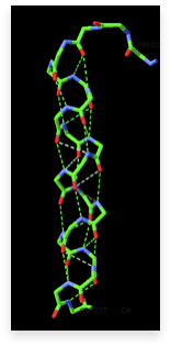
The most common type of secondary structure in proteins is the α-helix. Linus Pauling was the first to predict the existence of α-helices. The prediction was confirmed when the first three-dimensional structures of myoglobin and hemoglobin (Max Perutz and John Kendrew, 1962 Nobel Prize in Chemistry) were determined by X-ray crystallography. An example of an α-helix is shown in the image on the right. This type of protein structure representation is called “sticks representation”. In the image, only the main chain atoms of the polypeptide are shown connected by sticks, and the side chains are omitted. There are 3.6 residues/turn in an α-helix, meaning there is one residue every 100 degrees of rotation (360/3.6). Each residue is translated 1.5 Å along the helix axis, giving a vertical distance of 5.4 Å between structurally equivalent atoms in a helical turn (pitch of a turn). The repeating structural pattern in helices results from similar φ and ψ torsion. This is reflected in the clustering of the torsion angles within the helical region of the Ramachandran plot. In the section on the Ramachandran plot, we call these regions “energetically most favorable”. This means that the particular configuration of the polypeptide chain is such that main chain atoms are packed in an optimal and energetically favorable way, avoiding clashes (atoms coming too close to each other).
When looking at the helix in the image, notice how the carbonyl (C=O) oxygen atoms (shown in red) point in one direction towards the amide NH groups four residues away (i, i+4). Together, these groups form a hydrogen bond, one of the main forces in stabilizing protein secondary structures. The hydrogen bonds are shown as dashed lines.
The α-helix is not the only helical structure found in proteins. Other helical structures include the 310 helix, which is stabilized by hydrogen bonds of the type (i, i+3), and the π-helix, stabilized by hydrogen bonds of the type (i, i+5). Additionally, there is the left-handed L-α helix. The 310 helix has a smaller radius than the α-helix, while the π-helix has a larger radius. The first detailed analysis of the occurrence of the π-helix in proteins, based on entries from the Protein Data Bank (PDB), was conducted by Fodje and Al-Karadaghi, 2002. Generally, all these helical structures are relatively rare compared to the α-helix. In addition to these “simple” helical structures, there are also coiled-coil formations, where two or more α-helices intertwine to form higher-order helical structures.
β-Strands and the β-Sheet
The second major secondary structure element in proteins is the β-sheet. β-sheets consist of several β-strands, stretched segments of the polypeptide chain kept together by a network of hydrogen bonds between adjacent strands. An example of a β-sheet, with the stabilizing hydrogen bonds between adjacent strands (dotted lines), is shown in the image below (left):
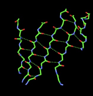
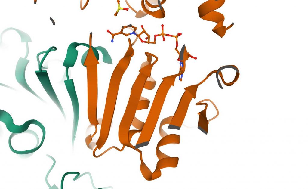
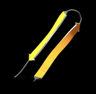
It is important to note that unlike in helices, in which the hydrogen bond donors and acceptors along the helix axis are separated by 4 residues, in β-sheets the hydrogen bond donors and acceptors belong to different strands and may be separated from each other by long segments of the amino acid sequence. In the upper image on the right, a ribbon presentation of a β-sheet is shown, this time in the context of the 3D structure to which it belongs (the coloring is according to secondary structure). Each β−strand is represented by an arrow, which defines its direction starting from the N- to the C-terminus. When the strand arrows point in the same direction, it is a parallel β-sheet (the PDB code for this structure is 2OHX).
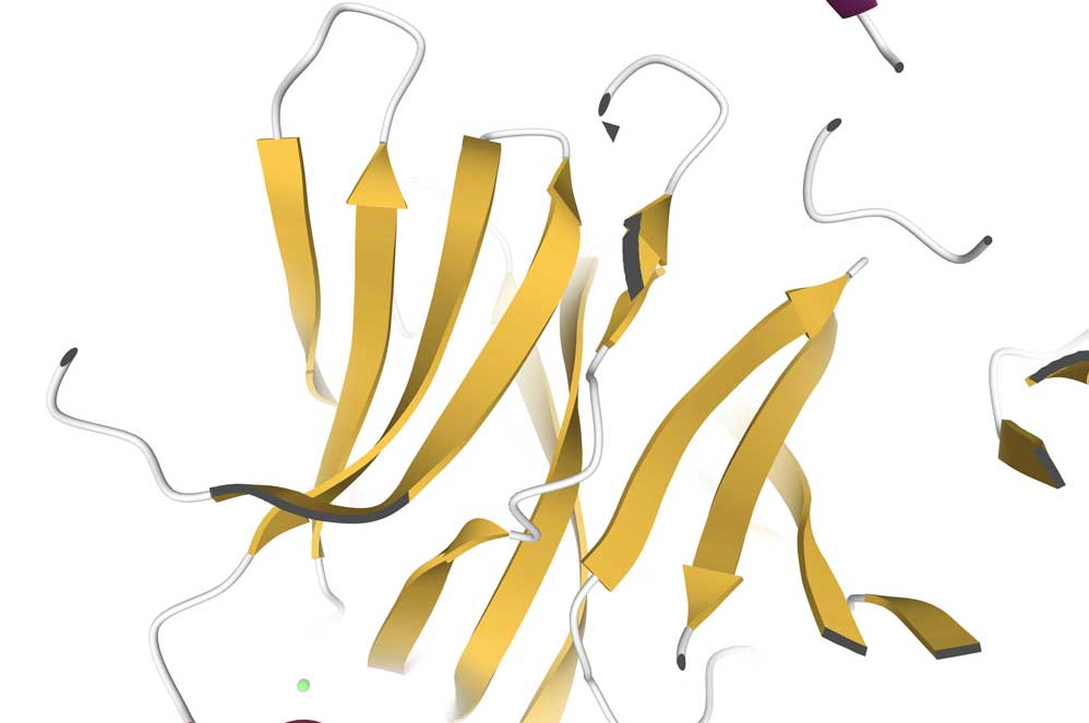
When the strands run in opposite directions (N-terminus to C-terminus vs. C-terminus to N-terminus), we get an anti-parallel β-sheet (PDB code is 1USR, Newcastle disease virus hemagglutinin-neuraminidase), images below:
Protein Structural Motifs
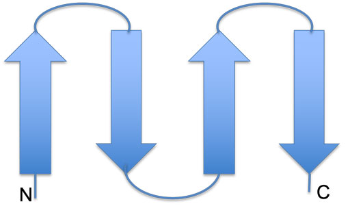
When linked together, secondary structure elements form structural motifs. For two antiparallel β-strands, such as shown in the figure above on the right, the structural motif is referred to as a β-hairpin. The loop connecting the two strands is known as a β-turn.
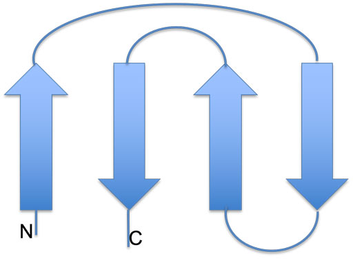
Short turns and longer loops are essential in protein 3D structures, and when they connect β-strands to β-strands, β-strands to α-helices, or α-helices to α-helices, they create various motifs. The β-hairpin mentioned above is just one example; others include the Greek key, helix-loop-helix, helix-loop-strand, and the zinc finger motifs. The image on the right shows a schematic presentation of the Greek key motifs, while on the left, two hairpins are connected into an anti-parallel β-sheet.
The amino acid sequences in loop regions are often highly variable within a protein family. Nevertheless, sometimes, when a loop has some specific function, for example, interaction with another protein, the sequence may be conserved. Loop length in organisms living at elevated temperatures (thermophilic organisms) is usually shorter than in proteins from lower-temperature family members. A shorter loop gives a protein additional stability at high temperatures, preventing its unfolding and denaturation. When making sequence alignment, a region with many insertions and deletions usually reveals the position of loops.
The following section will examine the next level of organization of protein structures – folds and domains.